Figure 7.
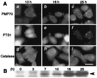
Kinetics of peroxisome biogenesis. (A) ZP119EG1 cells expressing EGFP-PTS1 were transfected with pUcD2Hyg⋅HsPEX19, then monitored by fluorescent microscope. (a–c) PMP70 was visualized by using rabbit anti-PMP70 antibody and Texas Red-labeled goat anti-rabbit IgG antibody. (d–f) EGFP-PTS1. (g–i) Catalase in other cells detected by anti-catalase antibody, as for PMP70. (a, d, and g) 10 h after transfection, (b, e, and h) 16 h. (c, f, and i), 25 h. Note that PMP70, but not EGFP-PTS1 and catalase, is already in numerous vesicular structures at 10 h (see text). (Bar = 20 μm.) (B) Expression of human Pex19p in ZP119. Pex19p was detected by immunoblotting HsPEX19-transfected ZP119 lysates (1.3 × 10⁵ cells at 0 h), at indicated time, where cell-doubling time was 22 h. Arrowhead, Pex19p.

