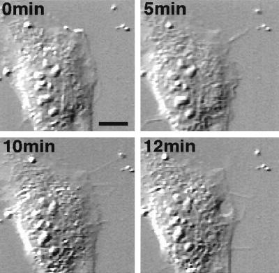
Figure 3.
Time-lapse images of a cell injected with V23RalA. V23RalA protein (1.6 mg/ml) was microinjected into serum-starved subconfluent fibroblasts, which were recorded under differential interference contrast microscopy (Nikon TE300). Images were collected with a charge-coupled device camera and time-lapse controller (ARGUS-20, Hamamatsu Photonics, Hamamatsu City, Japan), transferred to a Macintosh computer with a frame grabber (Scion, Frederick, MD), and processed by using National Institutes of Health image analysis. Frames at selected times after injection were shown. (Scale bar represents 10 μm.)

