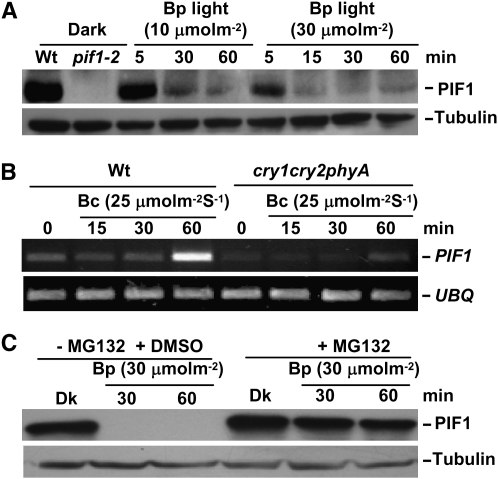
Figure 4.—
Blue light induces rapid degradation of PIF1 through the ubi/26S proteasomal pathway. (A) Native PIF1 is rapidly degraded after exposure to a pulse of blue (Bp) light conditions. Four-day-old dark-grown seedlings were exposed to Bp light (10 or 30 μmol m−2) and then incubated in the dark for the time indicated before harvesting for protein extraction. As controls, protein extracts from dark-grown wild-type and pif1 seedlings are included in the first two lanes, respectively. Approximately 30 μg of total protein in each lane were separated on an 8% polyacrylamide gel, transferred to PVDF membrane, and probed with anti-PIF1 antibody. A similar blot was probed with anti-tubulin antibody. The bands corresponding to PIF1 and tubulin are labeled. (B) PIF1 is slightly induced under blue light conditions. RT–PCR analyses are shown of PIF1 mRNA levels extracted from 4-day-old dark-grown seedlings or 4-day-old dark-grown seedlings exposed to continuous blue light (25 μmol m−2 sec−1) for the durations indicated. UBQ10 was used a control for the RT–PCR assays. (C) Blue light-induced degradation of PIF1 is mediated through the ubi/26S proteasomal pathway. Four-day-old dark-grown seedlings were pretreated with or without MG132 (30 μm) for 5 hr before being exposed to Bp light (30 μmol m−2) and then incubated in the dark for the durations indicated.

