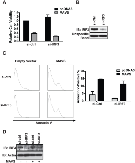
Figure 6. MAVS-induced apoptosis does not depend on IRF3.
(A) 1.0×10⁴ HEK293T cells were plated in 96-well plates, a pool of four siRNA targeting IRF3 or control siRNA were added to each well, 24 hours later 200 ng MAVS or empty vector plasmids were transfected into the cell. XTT assay was performed 48 hours thereafter. Error bars shown in the plot represents three biological replicates. (B) The same IRF3-targeting and non-targeting siRNA transfection reagents used in (A) were applied to 5.0×10⁵ cells, and all cells were lysed in RIPA buffer 48 hours post-transfection and subjected to Western blotting to confirm knockdown. (C) 5.0×10⁵ HEK293T cells were plated in 6-well plates and treated with IRF3 or control siRNAs, 24 hours later 3 µg of MAVS expression plasmid or empty vector plasmids were introduced into the cells. Half of the cells from each well were harvested 48 hours thereafter and stained with Annexin V. The plot represents two separate experiments and the percentages of Annexin V positive cells were averaged for quantification. (D) The other half of the cells described in (C) were lysed in RIPA buffer and subjected to western blot analyses to confirm IRF3 knockdown.

