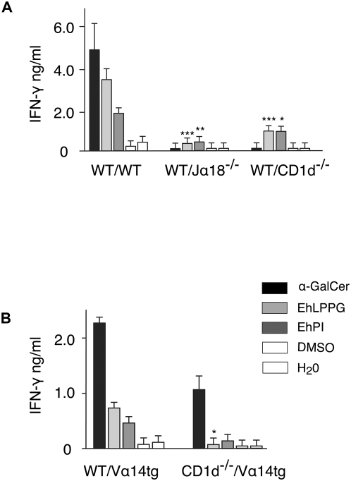
Figure 5. CD1d-restricted NKT cell activation by EhLPPG and EhPI.
(A) APC (5x10⁴) from WT mice were pulsed with either 2 µg of α-GalCer, EhLPPG or EhPI and incubated with 1x10⁵ lymphocytes of spleen cell preparations from Jα 18−/− lacking iNKT cells and CD1d−/− mice lacking iNKT and dNKT cells. IFN-γ secretion was measured by ELISA. Difference of IFN-γ production in EhLPPG/EhPI activated WT splenocytes *( p<0.05);**( p<0.005),***( p<0.001); ANOVA, Dunnett. (B) APC were generated from either WT or CD1d−/− mice, pulsed as described above and incubated with gradient purified liver lymphocytes from Vα14-transgenic mice. Liver iNKT cells were further purified by magnetic cells sorting. IFN-γ was assessed by ELISA. Difference of IFN-γ production in EhLPPG/EhPI activated WT splenocytes *( p<0.05); student t test. The results were obtained from three independent experiments.

