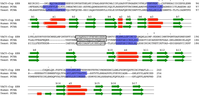
Figure 1. Comparative alignment between VACV G8R, human and yeast PCNA.
Alignment created using T-Coffee alignment algorithm, default parameters, with minor adjustments made by hand [20]. Green arrows represent beta-strands, and red cylinders represent alpha-helices. Black rounded box shows the interdomain connecting loop, blue shaded boxes represent the helices that contribute to DNA binding and dots represent identical residues between the 3 protein sequences. Residue marked with an asterisk is the conserved ubiquitylation/SUMOylation site. Secondary structure was derived directly from the crystal structures of the human and yeast PCNA proteins and the structural model of the VACV G8R protein.

