Figure 4.
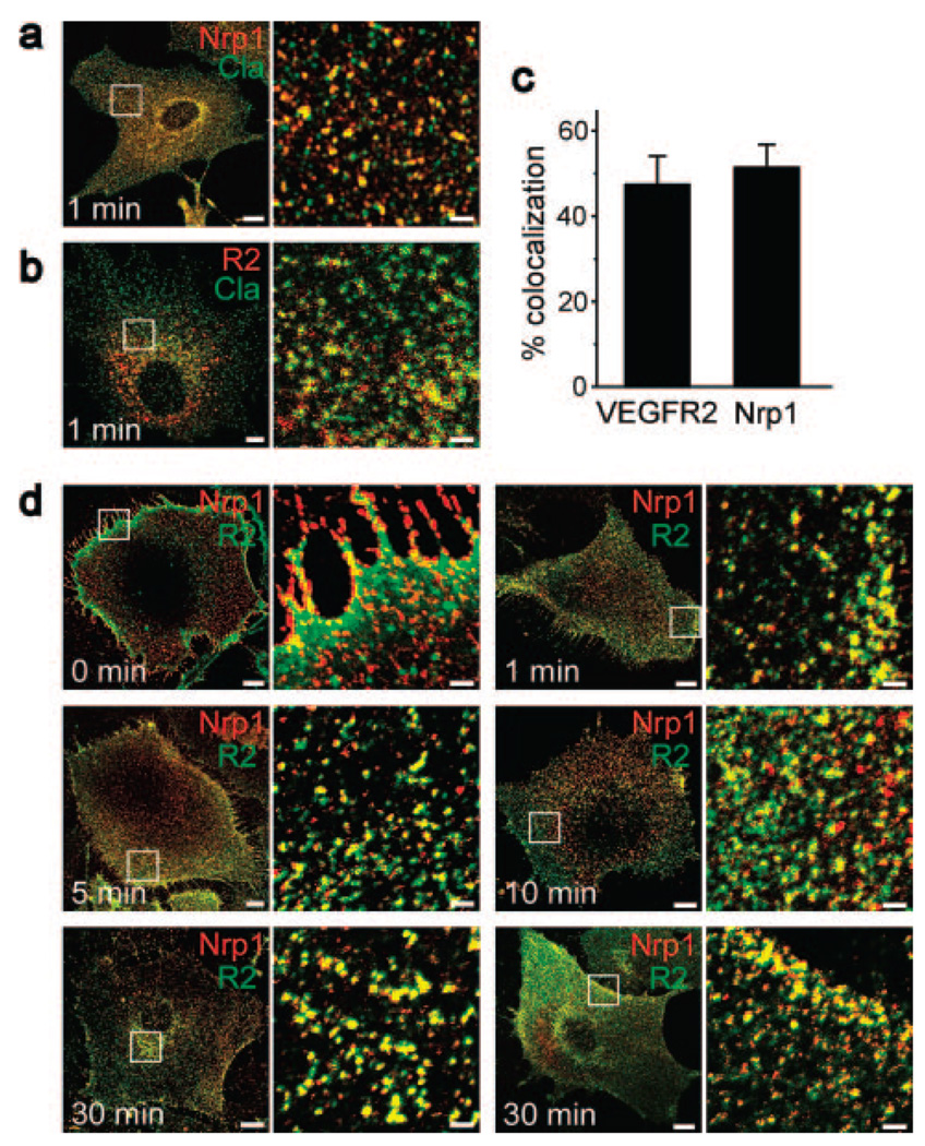
Nrp1 and VEGFR2 colocalized with clathrin and with each other during uptake in response to VEGF-A165. a and b, Mouse ECs fixed 1 minute after the start of uptake in response to VEGF-A165 (50 ng/mL), labeled with anti-Nrp1 (a) or anti-VEGFR2 (R2) (b) (both red) and with anti-clathrin (Cla) (green). Scale bars: 10 µm (2 µm in the magnified subfields). c, Extent of colocalization of Nrp1 or VEGFR2 with clathrin in VEGF-A165–treated ECs (n=3; mean±SD). d, Mouse ECs fixed at the indicated time points during uptake in response to VEGF-A165 (50 ng/mL), labeled with anti-Nrp1 (red) and anti-VEGFR2 (R2) (green). Scale bars: 10 µm (2 µm in the magnified subfields). The time course of Nrp1-VEGFR2 colocalization is shown in Figure 3b.

