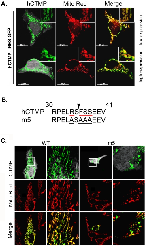
Figure 1. Interfering with CTMP maturation leads to swollen mitochondria.
(A) Full length human CTMP tagged with GFP was transfected into HeLa cells at low (upper panels) or high (lower panels) levels of expression. Mitochondria were visualized with Mitotracker Red. Merged fluorescence indicates CTMP-GFP expression in mitochondria. The round appearance of mitochondria is visible in cells with high levels of CTMP expression (lower panels). (B) Amino acid sequence of the R-2 predicted MPP cleavage site (indicated by the arrow) in human CTMP and a CTMP mutant (m5) in which R34, F36 and S37/38 have been mutated to alanine. (C) Twenty-four hours after transfection with CTMP-IRES-GFP or the m5 point mutant, HeLa cells were fixed and stained for CTMP and mitochondria as indicated. A detail of the squared area is shown in the right panel. Representative confocal pictures of three independent experiments are shown.

