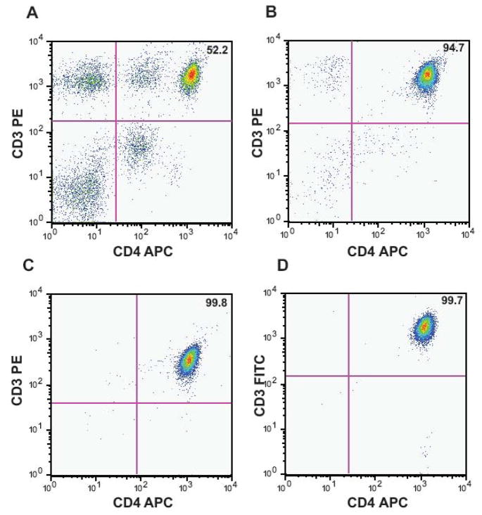
Figure 1. Isolation of CD3+CD4+ T cells by IMACS followed by FACS.
CD4+ T cells were purified from PBMC of healthy donors using negative selection by IMACS followed by one of two different FACS protocols, IMACS/FACS-1 and IMACS/FACS-2. IMACS-FACS-1 isolated CD3+CD4+ cells. IMACS/FACS-2 isolated CD4+ cells and excluded CD19+, CD123+, CD1c+, CD14+ and CD56+ cells. Following each purification step, cells were labeled with anti-CD3 and anti-CD4 mAbs and percent CD3+CD4+ cells was calculated in PBMC (52.2%, A), IMACS (94.7%, B) IMACS/FACS-1 (99.8%, C) and IMACS/FACS-2 (99.7%, D). One representative experiment of five using PBMC isolated from separate donors is shown.

