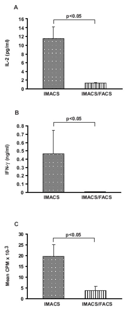
Figure 4. IMACS-CD4+ produce cytokines and proliferate in response to anti-CD3 mAb in absence of exogenously added costimulation.
CD4+ T cells were purified by IMACS (IMACS) or IMACS followed by FACS (IMACS/FACS) and cultured (105 cells/well) in anti-CD3 coated- flat-bottom 96 well plates (10 μg/ml). ( IL-2 was quantified in cell- A) free culture supernatants (18h) by ELISA. (B) IFN-γ was measured in cell-free culture supernatants (120h) by ELISA. (C) Proliferation was determined in 120h cultures by [3H] thymidine incorporation and results expressed as CPM. Mean values ± SEM of 5 experiments with separate donors are shown.

