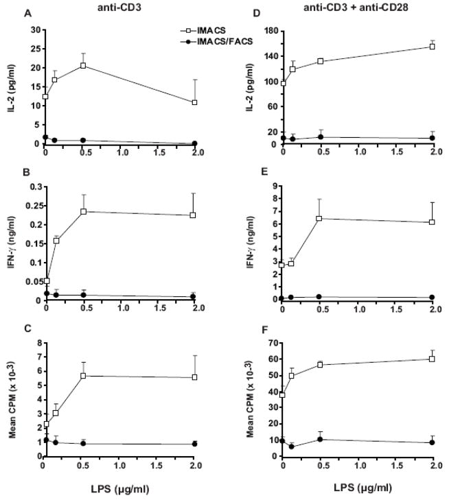
Figure 6. TLR-4 ligand LPS increases proliferation and cytokine secretion in IMACS- CD4+ but has no effect on IMACS/FACS-CD4+.
CD4 + T cells were purified by IMACS or IMACS/FACS and cultured (105 cells/well) in anti-CD3 coated- flat-bottom 96 well plates without (A–C) or with (D–F) soluble anti-CD28 (1 μg/ml) and different concentrations of LPS. (A, D) IL-2 was quantified in cell-free culture supernatants (18h) by ELISA. (B, E) IFN-γ was quantified in cell-free culture supernatants (72h) by ELISA. (C, F) Proliferation was measured at 72 hours by [3H]thymidine incorporation and results expressed as CPM. Mean values ± SD of triplicates from a representative experiment of two are shown.

