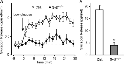
Figure 3. Impaired glucagon secretion in isolated synaptotagmin-7 KO mouse islets.
A, low glucose-induced glucagon secretion from isolated islets was measured in perifusion experiments at a glucose concentration of 10 mm (basal) or 1 mm (stimulatory). Arrow indicates switching from basal to stimulatory perifusion buffer. The perfusate was collected in 2 min intervals, and glucagon levels were determined by using RIA. Synaptotagmin-7 KO islets (Syt7−/−, filled circle) showed impaired glucagon secretion when compared with control (open circle). B, low glucose-stimulated glucagon secretion for the entire stimulation period in the perifusion experiments was lower in isolated islets from synaptotagmin-7 KO (grey bar) than from control (white bar). Glucagon secretion was calculated by integrating the area under each curve in A after baseline subtraction. Data are presented as means ±s.e.m., n= 7 for KO and 9 for control, **P < 0.01.

