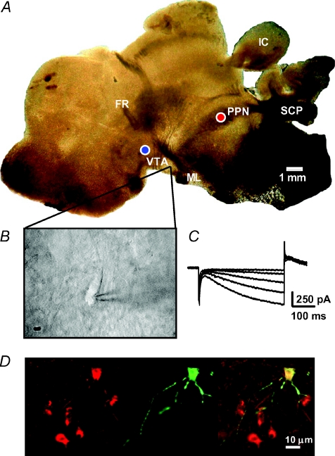
Figure 1. Identification of neurons in PPN-VTA brain slices.
A, photomicrograph showing a 280 μm thick parasagittal brain slice preparation containing the PPN and VTA as it appears in the brain slice recording chamber. The placement of stimulation and recording electrodes in the VTA (blue circle) and the PPN (red circle) was done using the superior cerebellar peduncle (SCP), and the fasciculus retroflexus (FR) as landmarks. Also aiding in the orientation were the medial lemniscus (ML) and inferior colliculus (IC). Note that in the brain slice the overlying cerebral cortical layers were removed during dissection (see supplemental file describing the blocking and slicing procedure). B, video microscopic image of a VTA neuron selected for recording. Neurons were visualized using a gradient contrast microscope and infrared illumination. C, inward currents in response to a series of negative voltage steps. Only neurons exhibiting large Ih, such as that shown in C, were included in this study. D, photomicrograph of a VTA neuron filled with biocytin (image at center) and counterstained for the dopamine transporter (image at left). At right is shown the superimposed images.

