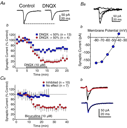
Figure 5. Identification of neurotransmitter receptors mediating PPN-evoked synaptic currents in VTA neurons.
Aa, PPN-evoked synaptic current that was representative of the majority of neurons in displaying partial sensitivity to a saturating concentration of the AMPA receptor antagonist DNQX (10 μm). b, mean time course illustrating 2 populations of PPN-evoked synaptic currents in VTA neurons; a majority inhibited less than 50% by DNQX (blue squares), and a minority inhibited greater than 50% (red circles). Ba, representative PPN-evoked synaptic currents remaining following DNQX application in a VTA neuron. The currents were evoked while the VTA neuron membrane potential was stepped from −40 mV to −80 mV (b). These synaptic currents remaining after DNQX application reversed near the Nernst-predicted reversal potential for Cl− (predicted: −46 mV, actual: −43 mV). The nAChR antagonists MLA (50 nm) and DHβE (1 μm), and the muscarinic cholinergic antagonist, atropine (1 μm) were also present in the aCSF during these experiments. Ca, mean time course of the effects of the GABAA antagonist bicuculline (10 μm) on PPN-evoked synaptic currents recorded in VTA neurons. Two populations of PPN-evoked synaptic currents were observed: those completely unresponsive to bicuculline (No effect, blue squares), and those inhibited by approximately 50% by bicuculline (Inhibited, red circles). b, representative signal averages of PPN-evoked synaptic currents that were sensitive (top, red line), and insensitive to bicuculline (bottom, blue line). Control traces are shown in black.

