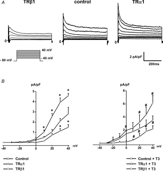
Figure 3. TRα1 increased native Ito in cardiomyocytes, while overexpression of TRβ1 resulted in a significant reduction of Ito.
Representative original current traces recorded in infected myocytes (A) and current density–voltage relations of each group (B) demonstrated that expression of exogenous TRα1 increased Ito current density which was even more pronounced upon T3 treatment. Conversely, overexpression of TRβ1 significantly suppressed Ito1 current size. Control cells were infected with adenoviral vectors expressing EGFP alone. *P < 0.05 versus control, #P < 0.05 compared with control+T3.

