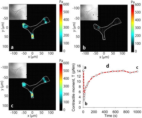
Figure 2. CMR measurements for a representative cell.
a, Traction map before cell stretch. b, Traction map measured immediately after an imposed homogeneous biaxial stretch of a 4 second stretch-unstretch maneuver with a peak strain amplitude of 10%. The cell tractions are markedly ablated. c, Traction map measured at 1000 seconds following stress cessation. Tractions have largely recovered to the baseline pre-stretch value measured in (a). d, The traction field can be used to compute the contractile moment, T, corresponding to an equivalent force dipole.[14] At the earliest measurable time point following stretch (b), the contractile moment was significantly reduced to 20% of its baseline value (a) followed by a slow recovery (c).

