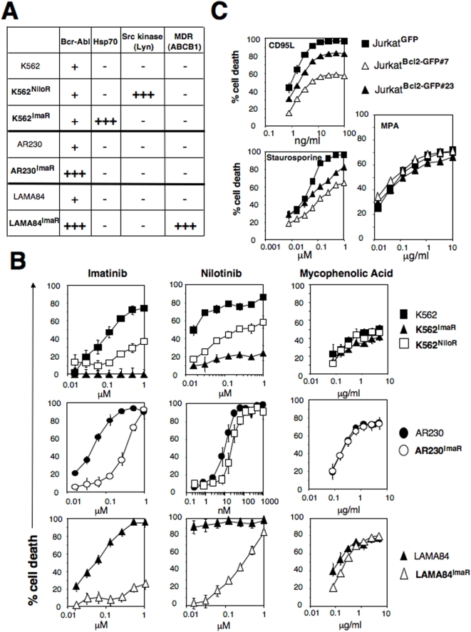
Figure 4. MPA mediates necrosis in malignant cells resistant to apoptosis.
(A) Molecular characterization of the imatinib and nilotinib-resistant CML cell lines. MDR: multidrug-resistance related protein. (−) indicates an expression level similar to the untreated cells, (+) refers to the ectopic expression of BCR-Abl and (+++) indicates the over-expression of the indicated protein. (B) Imatinib or nilotinib-resistant CML cells were incubated with the indicated concentrations of imatinib (left panel), nilotinib (middle panel) or MPA (right panel) for 48 hours. Cell death was measured by MTT assay. (C) Expression of Bcl2 (endogenous and GFP-Bcl2) was assessed in GFP- and GFP-Bcl2-expressing clones by immunoblot. The indicated cells were incubated for 24 hours with soluble CD95L, staurosporine or MPA for 48 hours. Total cell death was measured by MTT.

