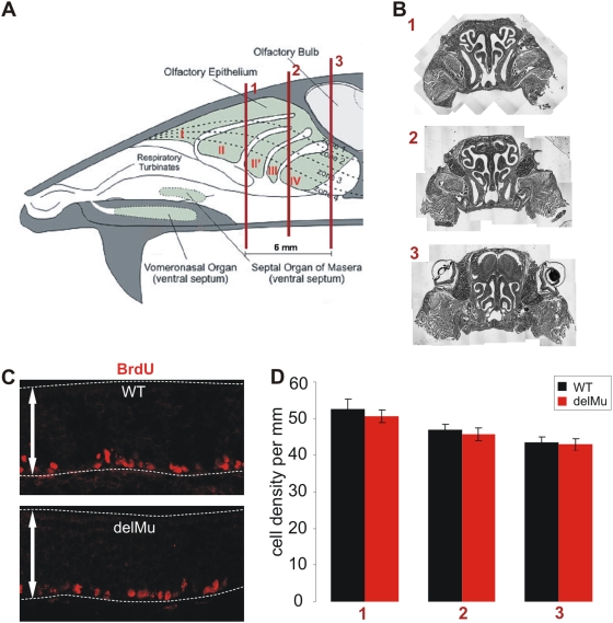
Figure 4. Quantification of BrdU-labeled basal cells in the olfactory epithelium of wild-type and eNOS deficient mice shows no difference in numbers of proliferating cells.
(A) Schematic drawing of a mouse head with the three coronal section levels (referred to as 1, 2 and 3) that were used for quantification of BrdU positive cells. Scheme adapted from A. Puche (www.apuche.org). (B) Reconstructions of coronal sections from these section levels and their respective localization (1, 2 and 3). (C) Exemplary anti-BrdU immunofluorescence from the nasal septum of wild-type (WT) and deletion mutant (delMu) mice. Dashed lines indicate the olfactory epithelium. Arrows represent 100 µm. (D) Cell densities of BrdU positive cells of the nasal septum of WT and delMu animals for all three section levels. Error bars indicate SEM.

