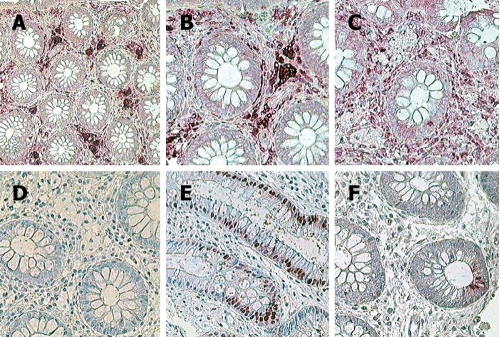
Figure 2.
Immunocytochemistry of CCC using monoclonal antibodies and APPAP method showing positive expression of mononuclear phagocytic cell line (A and B), negative T3 and T4 lymphocytes (C), negative neurofilament (D), Normal PCNA expression in cryptal epithelium (E), and normal mitotic activity of positive CD67 (F) (× 320).

