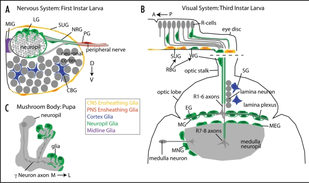
Figure 2.
Arrangement of glial types in the Drosophila nervous system. (A) Cross-section of a single hemi-segment of the embryonic ventral nerve chord. The midline is to the left. Embryonic and larval glia are: MIG, midline glia; LG, longitudinal glia; SUG, surface glia; NRG, nerve root glia; PG, peripheral glia; CBG, cell body glia. (B) Horizontal view of the eye disc and frontal view of the optic lobe of third instar larvae, depicting both the location and shapes of glial subtypes: retinal basal glia (RBG), which comprise surface glia (SUG) that ensheath the eye disc as a whole and wrapping glia (WG) that ensheath individual R cell bundles; SG, satellite glia; EG, epithelial glia; MG, marginal glia; MNG, medulla neuropil glia, MEG, medulla glia. (C) Mushroom body neuropil of the pupal CNS during axon pruning. An individual γ Neuron is highlighted. Upon an Ecdysone pulse, glial processes invade the neuropil and phagocytose degenerating axonal debris.

