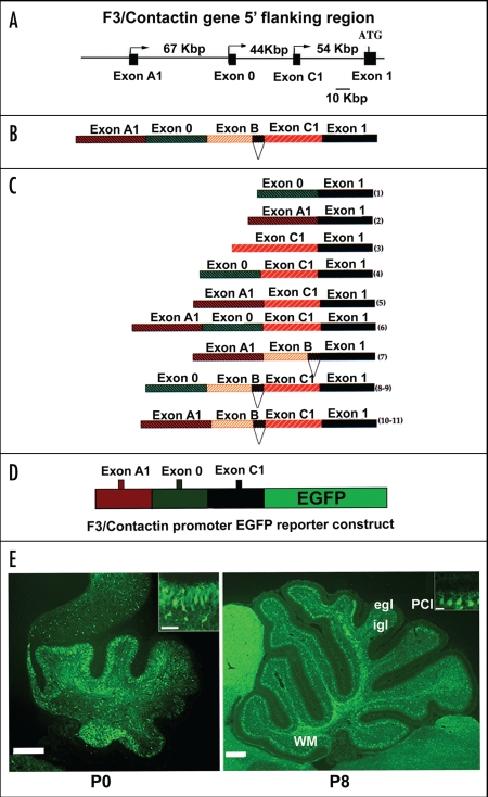
Figure 3.
Activation of the F3/Contactin promoter in developing mouse cerebellum. (A) Organization of the 5′ flanking region of the mouse F3/Contactin gene. In (B) the 5′ flanking exons are shown, which undergo complex splicing events (shown in C), resulting in a high level of complexity of the F3/Contactin mRNA (reviewed in ref. 66). (D and E) Map of the F3/Contactin promoter/EGFP reporter construct (D), and its expression in developing cerebellum (E). Transgene expression recapitulates the endogenous gene with an earlier activation on migrating granule cells (P0, see also inset) and subsequent expression on Purkinje neurons (P8, see also inset). Egl, external granular layer; Igl, inner granular layer; PCl, Purkinje cells layer; WM, white matter. Scale bars: P0, P8 = 200 µm (insets 20 µm).

