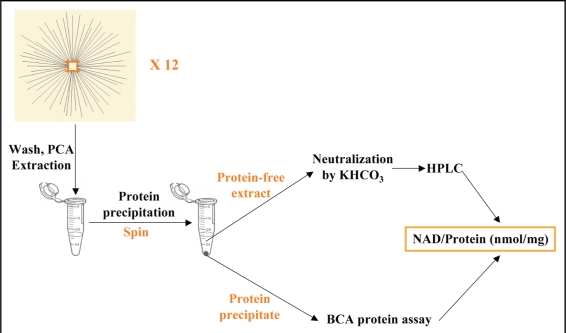
Figure 3.
The method used to measure axonal NAD+ level. Axons from 12 culture DRG explants were washed with PBS and extracted with perchloric acid. The precipitated protein was separated by centrifuge. The amount of the precipitated protein was measured by the modified BCA protein assay. The protein-free extract was neutralized and applied to HPLC. The axonal NAD+ level is normalized against the protein contents in the same sample and expressed as nmol/mg.

