Fig. 6.
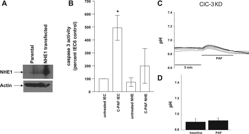
PAF-induced caspase 3 activity in IEC-6 cells is inhibited by Na/H exchanger 1 (NHE1) overexpression. A: immunoblot demonstrating overexpression of NHE1 in IEC-6 cells. B: caspase activity was determined in IEC-6 cells from cell lysates representing equal amounts of cellular protein using a fluorometric caspase 3 substrate in response to 1) basal conditions (no treatment), 2) PAF, 3) NHE1 transfection and 4) NHE1 transfection + PAF. Caspase activity was normalized to activity in untreated IEC-6 cells. The asterisk depicts statistical significance at P < 0.05 compared with basal conditions. C: representative tracing of pH measurements in ClC-3 knockdown IEC-6 cells by microfluorimetry using the pH-sensitive fluorescence dye SNARF and treatment with PAF 3 μM. In contrast to control cells (Figs. 1 and 2), no acidosis is seen in n > 3 separate experiments each representing minimum 7 cells/experiment, all with similar results. D: summary data from separate experiments with measurements taken from >7 cells/experiment indicating no PAF-induced acidosis in ClC-3 KD cells.

