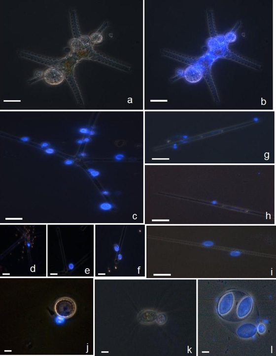
FIG. 2.
Examples of microscopic micrographs of phytoplankton eukaryotes with CFW-stained chytrid parasites, obtained via the fractionated community approach. Typical morphological taxonomic keys are visible under white light for host cell (i.e., the chlorophyte Staurastrum sp.) (a) and under UV light for the specific parasitic chytrids (b). Under UV light, chytrid epidemics were diagnosed for a diversified host populations, including both large size (e.g., the chlorophyte Staurastrum sp. [a and b] and the diatoms A. formosa [c to f] and Synedra sp. [g to i]) and small size (e.g., the diatom Cyclotella sp. [j] and the chlorophytes C. ciliata [k] and O. lacustris [l]) hosts. Multiple infectious chytrids are visible in most micrographs and different development stages as well, e.g., young sporangia with visible rhizoidal system (d and g), mature sporangia containing zoospores (e and h), a mature sporangium discharging its zoospore contents (f), and empty sporangia with chitinaceous wall visible (i). Scale bar, 10 μm.

