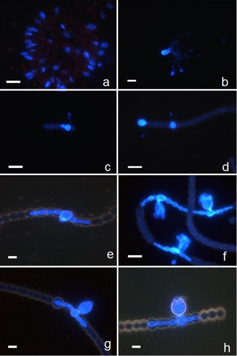
FIG. 3.
Examples of microscopic micrographs of phytoplankton prokaryotes (cyanobacteria) with CFW-stained chytrid parasites, obtained using the fractionated community approach. Typical morphological taxonomic characteristics are visible under white light for host cells (micrographs not shown) and under UV light for parasitic chytrids on their hosts identified as colonial Gomphosphaeria sp. (a) and Microcystis sp. (b) and as the filamentous Anabaena flosaquae (c to h). The branched rhizoidal system of the parasite is visible on Microcystis sp. (b). On A. flosaquae, young endobiotic thalli and encysted zoospores attached by long slender penetration tubes to the host are visible (c and d). In addition, tubular vegetative structure (e), mature sporangia of irregular pyriformic shape (f), and mature sporangia with protruding papilla for discharging zoospores (g and h) are also visible. Scale bar, 10 μm.

