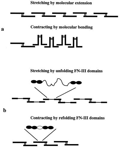
Figure 6.
Schematic diagram of possible mechanisms for stretching and contracting an FN matrix fibril. These diagrams illustrate single FN molecules; it is important to remember that the fluorescent FN fibrils are ten to hundreds of molecules thick, and hundreds of molecules in legnth. (a) In the contracted fibril the molecules are in a compact conformation, with bends between FN-III domains and stabilized by intramolecular bonds. Stretching the fibril breaks these bonds and extends the FN molecules. (b) The fibril could be stretched by unfolding FN-III domains and contracted by refolding the domains. The unfolded domain is 28.5 nm compared with the 3.5-nm length of the folded domain (13–15).

