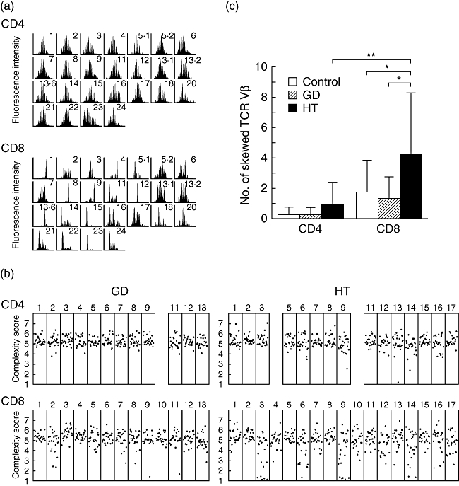
Fig. 1.

CDR3 spectratyping of T cell receptor (TCR) Vβ. (a) CDR3 size distribution. Each TCR Vβ fragment was amplified from cDNA with one of 25 Vβ-specific primers. The size distribution of polymerase chain reaction (PCR) products was determined by an automated sequencer and GeneScan software. Shown are the results of CDR3 size distribution in CD4+ and CD8+ T cells from patient 16 of Hashimoto's thyroiditis. (b) CDR3 complexity scores. Complexity scores for each TCR Vβ were shown. (c) Frequency of skewed TCR Vβ repertoire. Shown are the mean (±standard deviation) numbers of skewed TCR Vβ obtained from CD4+ and CD8+ T cells of the control and patients with Graves’ disease and with Hashimoto's thyroiditis. GD, Graves’ disease; HT, Hashimoto's thyroiditis. *P < 0·05; **P < 0·01.
