
Fig. 3.
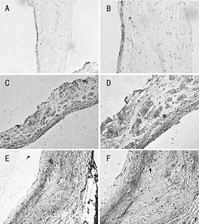
TN-C expression patterns in the normal, malformed and rheumatic valves. The normal (Fig. 2A and B), malformed (Fig. 2C and D) and rheumatic (Fig. 2E and F) valves were stained with polyclonal anti-TN-C antibodies as described in Materials and Methods. Magnification of 2A, 2C and 2E is 100-fold and that of 2B, 2D and 2F is 200-fold.