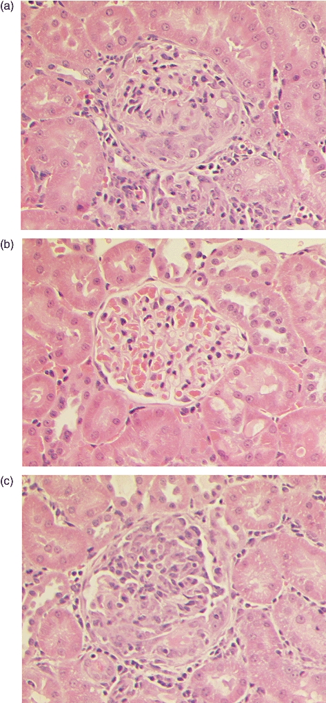
Fig. 5.

Light microscopy of kidney tissue at week 6 showing a representative example of: (a) marked segmental necrosis of the glomerular tuft with crescent formation in a positive control Wistar Kyoto (WKY) rat immunized with alpha 3 chain of type IV collagen [α3(IV)NC1]; (b) normal glomerular architecture in a negative control WKY rat given Freund's complete adjuvant (FCA) alone; and (c) severe crescentic glomerulonephritis in a WKY rat immunized with peptide pCol(24–38). Magnification ×300.
