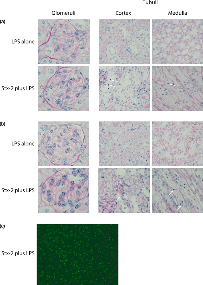
Fig. 1.

Renal histology in mice treated with Shiga toxin (Stx-2) and lipopolysaccharide (LPS). Light microscopic appearances of periodic acid-Schiff (PAS)-stained renal sections from (a) wild-type and (b) Cfh+/− animals treated with either LPS alone or 200 ng Stx-2 in combination with LPS. Representative glomerular, cortical and medullary tubular changes are shown. Arrows indicate examples of apoptotic tubular cells. Equivalent histological changes were seen in the tubules of mice treated with 50 ng Stx-2 in combination with LPS (data not shown). Original magnification was 40× for tubular and 100× for glomerular images. (c) Representative image of medullary terminal deoxynucleotidyl transferase-mediated dUTP-biotin nick end labelling (TUNEL) staining in wild-type mouse treated with Stx-2 and LPS. Equivalent staining was seen in Cfh+/− animals treated with Stx-2 and LPS.
