
Figure 2.
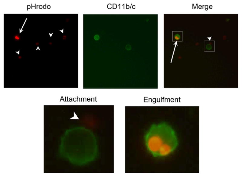
Fluorescent intensity of pHrodo-SE-labeled apoptotic thymocytes increases after engulfment by macrophages. Splenic macrophages were labeled with FITC-anti-CD11b/c (OX42) and thymocytes with pHrodo-SE (0.02 μg/ml). Cells were coincubated for 60 min, collected and fixed with 1%PFA prior to fluorescent microscopy. Top three panels show lymphocytes in red (pHrodo-SE+), macrophages in green (CD11b/c) and a merged image, of the same area to the right. Boxes in the merged image indicate the 10× magnification shown below. Arrowheads indicate free floating and attached apoptotic thymocytes and arrows indicate engulfed apoptotic cells. Magnification: 400×.