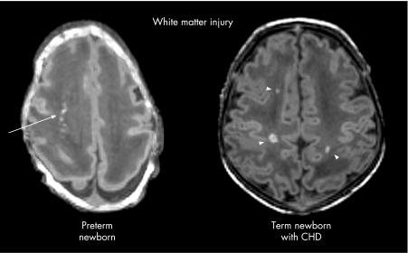
Figure 1 White matter injury in a premature newborn born at 28 weeks' gestational age and in a term newborn with congenital heart disease, both scanned at 2 weeks of life. The axial images from the spoiled gradient echo volumetric scans show several foci of T1 hyperintensity in the periventricular white matter of the preterm newborn (arrow) and of the term newborn with heart disease (arrowheads).

An official website of the United States government
Here's how you know
Official websites use .gov
A
.gov website belongs to an official
government organization in the United States.
Secure .gov websites use HTTPS
A lock (
) or https:// means you've safely
connected to the .gov website. Share sensitive
information only on official, secure websites.
