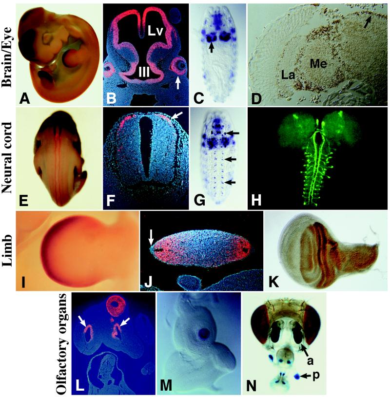
Figure 2.
Comparison of ap and mLhx2 expression patterns. (A) mLhx2 expression in the forebrain and limbs of an E11.5 mouse embryo. (B) At this stage, mLhx2 is expressed in the walls of the lateral ventricles (Lv) and third ventricle (III) of the brain. In the eyes, mLhx2 is expressed in the future nervous layer of the retina (arrow) and in the optic stalk (not shown). (C) ap expression in the brain hemispheres (arrow) of a stage 15 fly embryo. (D) In the adult fly, Ap is immunodetected in the lamina (La) and medulla (Me) of the optic lobe and in the central brain (arrow). (E and F) At E11.5, mLhx2 is expressed along the neural tube (E) in a group of dorsal commissural interneurons (arrow in F). (G) ap expression in the VNC (arrows) of a stage 15 fly embryo. Out of focus, expression is also evident in the brain hemispheres and muscles of the body wall and pharynx. (H) Drosophila larval central nervous system showing expression of a UAS:tau-GFP responder driven by the ap-VNC enhancer. Note the axonal projections of ap-expressing interneurons along ascending longitudinal tracts. (I and J) mLhx2 expression in E11.5 mouse limbs. Label is detected in the mesenchyme, in a region roughly corresponding to the progress zone (I). In cross-sections, mLhx2 is observed in both dorsal (up) and ventral (down) regions of the limb and is excluded from the apical ectodermal ridge (arrow in J). (K) Ap immunodetection in the dorsal compartment of a Drosophila wing imaginal disc. (L) Section from an E11.5 mouse embryo showing mLhx2 expression in the olfactory epithelium surrounding the nasal pits (arrows). (M) ap expression in the center of a Drosophila antennal disc. (N) X-Gal stain of a Drosophila adult head carrying the enhancer detector aprk568, which expresses lacZ in an ap-like fashion. Note lacZ expression in the fly olfactory organs: the antenna (a) and the palpus (p).

