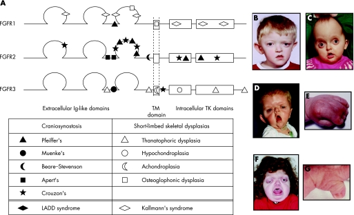
Figure 3 Fibroblast growth factor receptor (FGFR) structure and disease. (A) Structure of FGFR1, FGFR2 and FGFR3 proteins. Positions of some of the common mutations in different diseases are shown. Ig, immunoglobulin; TM, transmembrane; TK, tyrosine kinase. (B–G) Dysmorphic facial features, in some cases associated with limb malformations, in patients with Muenke's syndrome and right unicoronal craniosynostosis (B), Crouzon's syndrome (C), Apert's syndrome (face (D) and hand (E)) and Pfeiffer's syndrome (face (F) and thumb (G)). Parental/guardian informed consent was obtained for the publication of these figures.

An official website of the United States government
Here's how you know
Official websites use .gov
A
.gov website belongs to an official
government organization in the United States.
Secure .gov websites use HTTPS
A lock (
) or https:// means you've safely
connected to the .gov website. Share sensitive
information only on official, secure websites.
