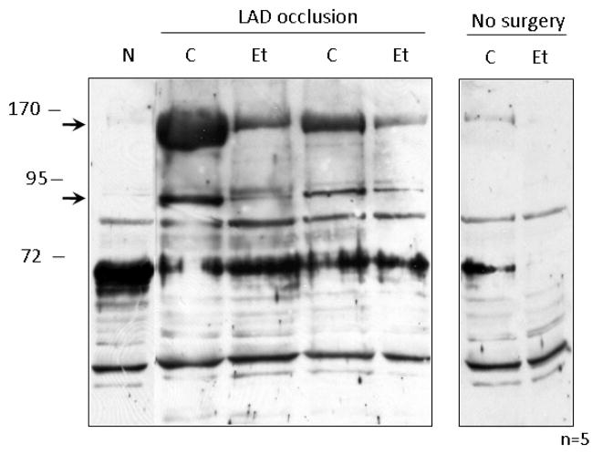
Figure 4. Ethanol treatment reduces HNE protein-adduct formation following I/R.
Mitochondrial protein was isolated from the left ventricles of animals that were administered 0.5 g/kg of ethanol 60 minutes prior to LAD occlusion (Et) and compared to animals that were not treated with ethanol (C) and animals which did not undergo LAD occlusion (N). The right panel shows basal levels of HNE formation in cardiac mitochondria that were not subjected to surgery. Following homogenization, mitochondrial lysate was analyzed by Western blot utilizing antibodies which recognize HNE protein adducts. Two protein bands which showed a reduction upon in HNE-adduct formation upon ethanol treatment are denoted by arrows.

