
Figure 3.
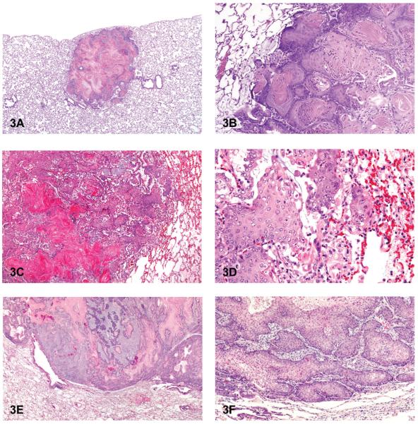
Cystic keratinizing epithelioma and squamous cell carcinoma. (A) Cystic keratinizing epithelioma. (B) Higher magnification of 3A. Note peripheral “cobblestoned” appearance of tumor cells and multiple layers of squamous cells surrounding central areas of keratin and inflammation. (C) Squamous cell carcinoma. (D) Higher magnification of 3C. Note nests of neoplastic squamous epithelial cells and focal areas of hemorrhage and inflammation. (E) Squamous cell carcinoma with cords of invasive tumor cells. (F) Higher magnification of 3E. Note nests and cords of neoplastic squamous cells. H & E. A, B: Sprague-Dawley (SD) rat, HF, PCB 126/PCB 118; C, D: SD rat, HF, PCB 126(3,3,′4,4,′5.5′-pentachlorophenyl); E, F: SD rat, MF, PCB 126/PCB 153.