
Figure 6.
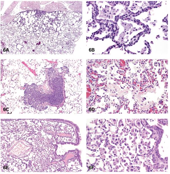
Hyperplasia and inflammation. (A) Hyperplasia with thickening of the alveolar septa. (B) Higher magnification of hyperplasia. Note maintenance of alveolar septal architecture, with no compression and lack of cellular or nuclear atypia of hyperplastic alveolar cells. (C) Focal infiltrate of acute inflammatory cells. (D) Infiltrates of mixed inflammatory cells. (E) Infiltrates of histiocytes. (F) Higher magnification of Figure 6E. Note alveolar sacs filled with numerous histiocytes. H & E. A: F344/N, MF, gallium arsenide; B: F344/N, HF, gallium arsenide; C: B6 mouse, HM, 2,3-dibromo-1-propanol; D: F344/N, rat, HF, hexachloropentadiene; E, F: B6 mouse, HM, gallium arsenide.