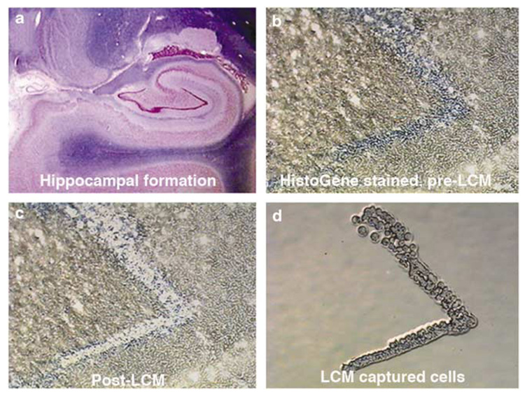
Figure 3.
LCM is used to collect dentate neurons from the human hippocampus (shown in lower power, top left) (a). The top right micrograph (b) is an enlargement that shows the blue-stained dentate neurons before LCM. In the lower right (d), the LCM captured neurons are isolated and gene expression is measured in cDNA produced from their amplified RNA. The ‘hole’ after LCM can be seen in the lower left micrograph (c).

