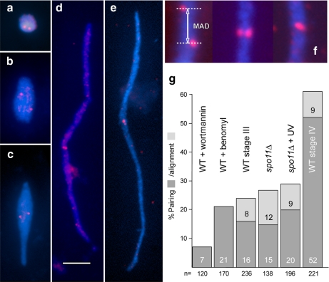
Figure 3.
Examples of meiotic MICs (a, stage I; b, stage II; c, stage III; d and e, stage IV) with FISH signals (red) representing unpaired loci in a–d and paired loci in e. The FISH probe (see Supplemental Figure S1) was selected so that it localizes to the middle of crescents. Because in the mature crescent (stage IV) all telomeres congress on one end (Mochizuki et al., 2008) and the centromeres on the opposite end (see Figure 2a), a locus found in the middle of a crescent must be situated in an intercalary position of a chromosome arm. This region has the advantage that its pairing behavior is largely independent of seeming pairing conferred by the clustering of centromeres and telomeres. Carnoy fixation removes MICs from the cells, and they become flattened and straightened out on the slide, which facilitates the quantitative evaluation of FISH signals. Bar, 5 μm. (f) Examples of misaligned, aligned, and paired loci within crescents. The misalignment distance (see text) was measured as indicated (MAD). (g) Pairing and alignment of FISH signals at different MIC elongation states. Pairing is infrequent in enlarged but round MICs of wortmannin-treated wild type (WT). After benomyl treatment, paring of the intercalary locus is only slightly increased despite centromeres becoming clustered in one spot at the periphery of the nucleus (see Figure 2g). The concept of aligned signals as used here (f) is not applicable to round nuclei. Also in slightly elongated nuclei (spo11Δ and WT stage III) pairing is still low. Notably, despite full nuclear elongation in the UV-treated spo11Δ mutant (see Figure 4), pairing does not reach the level of WT stage IV (n = number of nuclei inspected).

