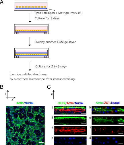
Figure 1.
HPPL form tubular structures in sandwich culture. (A) Schematic view of sandwich culture. HPPL were plated on gel containing type I collagen and Matrigel. After HPPL form a monolayer during 2 d of incubation, another layer of gel was overlaid on the monolayer. After additional 2 or 3 d of incubation, cells were fixed and examined with a confocal microscope after immunostaining. (B) A low-magnification image of sandwich culture stained with AlexaFluor 488–conjugated phalloidin and Hoechst34580. A tubular network was visualized as the area surrounded by thick F-actin bundles. Scale bar, 100 μm. (C) Vertical sections of sandwich culture. Cultures were stained with anti-cytokeratin 19 (CK19) antibodies and phalloidin (1–4) or phalloidin and anti-ZO1 antibodies (5–8). CK19+ cells formed the apical lumen surrounded by F-actin bundles. Formation of tight junction was visualized with ZO1 staining. Scale bars, 20 μm.

