Fig. 5.
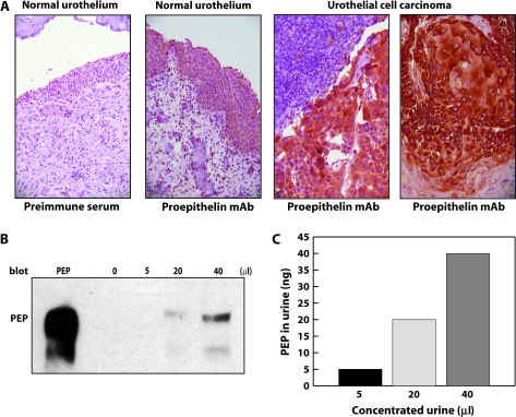
Proepithelin detection in bladder cancer tissue specimens and urine samples. (A) Formalin-fixed, paraffin-embedded sections of non-malignant human bladder and bladder cancer tissue were deparaffinated in preparation for antigen retrieval by heating in a microwave oven for 10 min. Slides were cooled, rinsed and immunostained using a monoclonal antibody raised against human proepithelin (A&G Pharmaceutical) and visualized with an avidin–biotin method. From left to right: negative control normal urothelium stained with preimmune serum (×200), normal urothelium expressing proepithelin (×200), high levels of proepithelin are detected in invasive urothelial cancer (×400) and in area of squamous differentiation (×400). (B) Random and unidentified urine samples from normal individuals were pooled and then concentrated (10 ml ⇒ 200 μl) using Amicon Ultra Centrifugal Filter Devices at 4000 r.p.m. for 20 min (Millipore's Corporate Headquarters, Billerica, MA, 10 K). Proepithelin levels were determined by immunoblot using anti-proepithelin polyclonal antibodies (Zymed Laboratories, Inc., South San Francisco, CA). Recombinant human proepithelin (300 ng) was loaded as control (PEP). Blot is representative of two independent experiments. (C) The amount of proepithelin present in urine was calculated by ELISA using a proepithelin ELISA kit (ALPCO Diagnostics).

