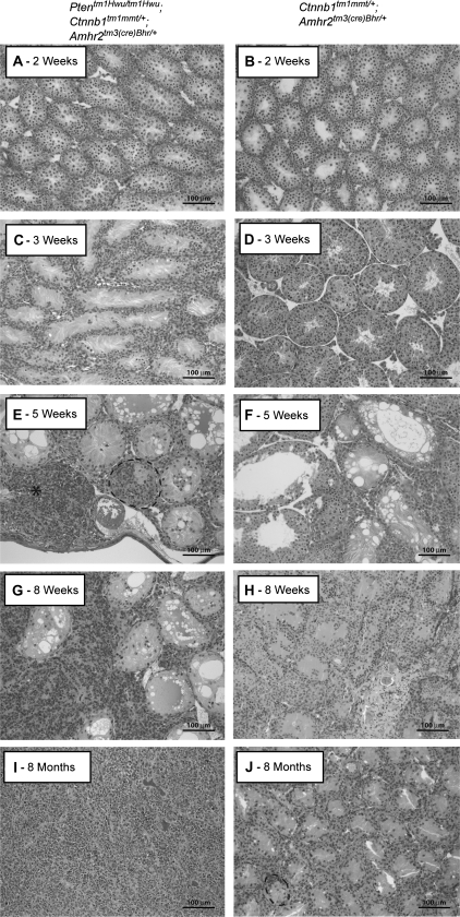
Fig. 1.
Formation of sex cord tumors in Ptentm1Hwu/tm1Hwu;Ctnnb1tm1Mmt/+;Amhr2tm3(cre)Bhr/+ mice. (A, C, E, G and I) Photomicrographs demonstrating progressive tumor formation and degeneration of the seminiferous tubules in Ptentm1Hwu/tm1Hwu;Ctnnb1tm1Mmt/+;Amhr2tm3(cre)Bhr/+ mice, compared with (B, D, F, H and J) Ctnnb1tm1Mmt/+;Amhr2tm3(cre)Bhr/+ mice at 2 weeks to 8 months of age. Multilayered, focal accumulations of cells in some seminiferous tubules are circumscribed with black dotted lines. A microtumor is indicated with an asterisk. Original magnification, ×200.

