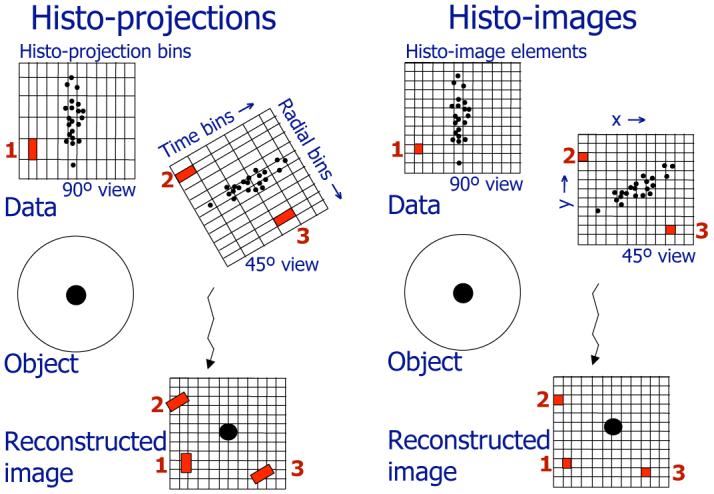
Fig. 1.

Comparison of the data formats for binned TOF data (histo-projections for 45° and 90° views - left) and the DIRECT approach (histo-images for 45° and 90° views - right) — Histo-projections can be viewed as an extension of individual non-TOF projections into TOF directions (time bins), and their sampling intervals relate to the projection geometry and timing resolution. Acquired events are first histogrammed into the histo-projection bins; during the reconstruction process individual histo-projection bins are then repetitively traced through reconstructed image voxels (examples for 3 bins - 1, 2, 3 - are shown). In the DIRECT approach histo-images are defined by the geometry and desired sampling of the reconstructed image. Acquired events and correction factors are directly placed into the image resolution elements of individual histo-images (one histo-image per each view) having a one-to-one correspondence with the reconstructed image voxels. All reconstruction and data correction procedures are done directly, and very efficiently, in image space.
