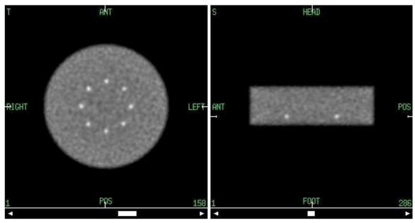
Fig. 9.

Transverse (left) and sagittal (right) slices of the short phantom (11 cm) with abrupt truncation of attenuation and emission activity at approximately 1/4 of the axial FOV (from both sides) of the simulated scanner. The edges of the 10-mm spherical lesions are located axially 15 mm from the phantom edge (phantom diameter 35 cm, 600-ps TOF resolution, DIRECT reconstruction using RAMLA with resolution modeling, 40×3 views, 4-mm voxels, 15 iterations).
