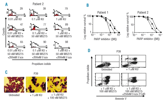
Figure 3.
The effect on leukemic cells of the PARP inhibitor, KU, in combination with non-cytotoxic concentrations of 5’-aza-2’-dCR and/or HDAC inhibitor. (A) The effect of the PARP inhibitor, KU, in combination with the HDAC inhibitor, MS275, and with 5’-aza-2’-dCR on primary AML cells. KU was added at variable concentrations to cells from AML patient # 2 continuously for 5 days with 50 nM MS275 and 200 nM 5’-aza-2’-dCR or KU alone and the cells were then analyzed by flow cytometry. The apoptotic index (sub-G1 population as a fraction of sub-G1 + G1 populations) is shown in the right inset. (B) Trypan blue exclusion assays were used to determine cell sur vival in primary AML cells from patient # 2 exposed to KU, MS275 and 5’-aza-2’-dCR. Cells from AML patients # 1 and # 2 were exposed continuously for 5 days to 50 nM MS275 and 200 nM 5’-aza-2’-dCR with varying concentrations of KU. KU + MS275 (triangles), 5’-aza-2’-dCR + KU + MS275 (diamonds), and KU alone (squares). (C) P39 cells were cultured in 1 μM KU, 1 μM KU + 100 nM MS275 or left untreated for 7 days. At 7 days cells were cytospun onto slides and stained with May-Grünwald stain. Images were taken at X40 magnification. (D) 1 μM KU, 100 nM MS275 and 250 nM 5’-aza-2CdR were added as indicated to P39 cells for 7 days before the cells were treated with annexin V-fluorescein isothiocyanate (FL-1, X-axis) and propidium iodide (FL-2, Y-axis).

