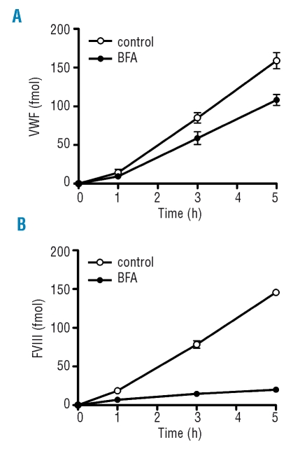
Figure 5.
Quantitative analysis of the secretion pathways of FVIII and VWF in FVIII-GFP transduced blood outgrowth endothelial cells. Release of FVIII from passage 12 FVIII-GFP transduced BOECs was analyzed over a 5-hour period in the presence of 5 μM BFA. (A) VWF antigen in the conditioned medium was quantified by ELISA. Values represent the mean ± SD of three experiments. Open circles (○) represent controls, closed circles (●) represent secretion of VWF in the presence of BFA. (B) FVIII antigen in the conditioned medium was quantified by ELISA. Values represent the mean ± SD of three experiments. Open circles (○) represent controls, closed circles (●) represent secretion of FVIII in the presence of BFA.

