
Figure 5. Immunocytochemical staining for Ang(1−7) and ACE2 in the left ventricle of the mouse heart.
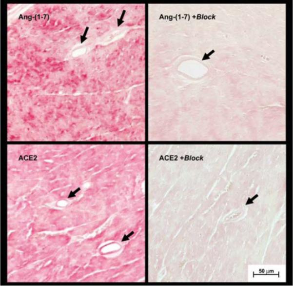
Sections of the left ventricle (5 μm) from the heart of wild-type mice were reacted with polyclonal antibodies against Ang(1−7) and ACE2. In the top left panel, Ang (1−7) staining is evident throughout the field, which is primarily composed of cardiomyocytes. In the bottom left panel, ACE2 staining was evident throughout the field and in coronary vessels. Pre-incubation of primary antibodies with Ang(1−7) (top right panel) or recombinant ACE2 (bottom right panel) essentially abolished the immunocytochemical signals. Data are representative of tissues from three animals. Arrows indicate coronary vessels and the scale bar represents 50 μm.