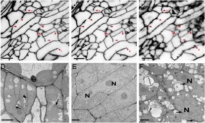
Figure 6.
Aberrant cell walls and two nuclei in one cell in gsl8-4 mutants. A to C, Cell wall stubs in epidermal cells visualized using propidium iodide staining and confocal microscopy. Optical sections from outer (A), middle (B), and bottom (C) parts of the same cells are shown. All propidium iodide images have inverted gray scales. The other five gsl8 mutant alleles showed similar results to those of gsl8-4. D to F, TEM of cotyledons (D) and root tips (E) from a 3-d-old gsl8-4 mutant and of a gsl8-4 embryo (F). Arrowheads and arrows indicate cell wall stubs. N, Nucleus. Bars = 10 μm in A to C and 2 μm in D to F. [See online article for color version of this figure.]

