Figure 5.
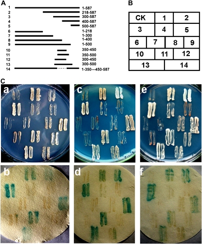
Transcription activation analysis of OsMYB3R-2 protein. A, Different pGBKT7-OsMYB3R-2 vector constructs. The truncated cDNA fragments of OsMYB3R-2 were sequenced and inserted into the NdeI-PstI sites, with ATG added at the end of the NdeI site in every forward primer. For numbers at left, 1 represents full-length OsMYB3R-2 protein and 2 to 14 represent different truncated OsMYB3R-2 protein fragments; numbers at right represent the positions of different truncated OsMYB3R-2 protein fragments. The broken line represents the deleted fragment (amino acids 351 to 449) of OsMYB3R-2. The transcription activation of OsMYB3R-2 was confirmed twice. B, The corresponding positions of transformed yeast thalli daubed on the plates. CK, pGBKT7 vector used as a control. C, a, The transformed yeast thalli grew on the SD/-His/-Trp plates with solid SD medium. C, b, X-Gal activation detection of transformed yeast thalli on the SD/-His/-Trp plates with solid SD medium shown in C, a. C, c, The transformed yeast thalli grew on the SD/-Ade/-Trp plates with solid SD medium. C, d, X-Gal activation detection of transformed yeast thalli on the SD/-Ade/-Trp plates with solid SD medium shown in C, c. C, e, The transformed yeast thalli grew on the SD/-His/-Ade/-Trp plates with solid SD medium. C, f, X-Gal activation detection of transformed yeast thalli on the SD/-His/-Ade/-Trp plates with solid SD medium shown in C, e.

