Figure 3.
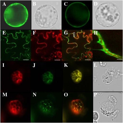
LTPG1:EYFP is localized in the plasma membrane of the Arabidopsis protoplast and tobacco epidermal cells. Arabidopsis protoplasts (A–D and I–P) and tobacco epidermis (E–H) transformed with each construct were visualized with a fluorescence microscope (Nikon Eclipse TE2000-U) and a laser confocal scanning microscope (TCS SP5 AOB5/Tandem; Leica), respectively. A and B, Control, p35S-EYFP-Flag/Strep plasmid. C to E, LTPG1:EYFP construct. F, Propidium iodide staining to visualize the cell wall. G, Merged images from E and F. H, Magnification of the inset box in G. I and M, LTPG1:RFPΔ48 construct. J, Bip:GFP construct. K, Merged images from I and J. N, ST:GFP construct. O, Merged images from M and N. Images were acquired through a GFP filter (A, C, E, J, and N), an RFP filter (F, I, and M), or a bright fields (B, D, L, and P). Bars = 25 μm (E–G) and 2.5 μm (H).

